
Author: Moorhouse Anna
Institution: English and Cell and Molecular Biology
Date: November 2005
"The event on which this fiction is founded has been supposed, by Dr. Darwin and some of the physiological writers of Germany, as not of impossible occurrence."
- P.B. Shelley, 1818
Percy Shelley's introduction to his wife Mary Shelley's novel Frankenstein: The Modern Prometheus, voices a belief that is prevalent in our society even now: in science, nothing is truly impossible.
In Shelley's novel, Dr. Frankenstein created life from death by reanimating the mismatched remains of cadavers using an electric current; however, the implicit moral in the story, that man cannot hope to control the creative powers of nature, suggests that toying with those powers will always end in disaster. Interestingly, it was the work of ambitious scientists like Dr. Erasmus Darwin, the grandfather of Charles Darwin, that is said to have first inspired Shelley and to have captured the collective imagination of her reading public.
Current research into tissue engineering and the media-push that has come with each new advance in the field have had no less affect on our own wildest dreams. If we are to believe every announcement that the cure for diabetes is only a step away and that laboratories across North America are already churning out new livers and skin grafts for everyone, why are we still waiting? A better question to ask is: what are scientists actually capable of creating right now, and how far do we still need to go?
'Dem Bones
Many people have hobbled into an emergency room sporting a newly broken leg or fractured wrist. With serious bone breaks, orthopedic surgeons typically remove small pieces of bone from a patient's rib or hip and fuse them to the broken bone in order to repair the damage. This procedure is very painful and can often produce major complications later on.
Every bone in our body is a dynamic piece of machinery, continually growing and reshaping its contours; however, the cellular processes in charge of stimulating bone growth are horrifically complex and intimately linked to stem cell research. V. Prasad Shastri, an assistant professor of biomedical engineering at Vanderbilt University, and his research team have been trying to harness these processes in order to understand how bones can be made to grow on demand.
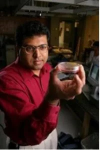
Figure 1. Prasad Shastri holding a petri dish containing bone cells. Photo courtesy Daniel Dubois.
In a paper published in August 2005, Shastri and his team describe what they have named an "in vivo bioreactor": a bone-making procedure using mature rabbits. They hollowed out a special zone on the surface of a healthy bone and waited while the rabbit's own wound-healing mechanisms worked. Six weeks after his team began their first trial, Shastri and his colleagues found that the bone site was full, and they harvested the new bone by detaching it from the old bone before the two fused.
"We have shown that we can grow predictable volumes of bone on demand," says Shastri. "And we did so by persuading the body to do what it already knows how to do."
Most long bones in humans and rabbits are covered by a thin outer layer known as the periosteum. While the outside of the layer is tough and fibrous, the inside is covered with pluripotent cells -- stem cells that are able to differentiate into many different types of tissues within the body -- in this case, skeletal tissues. The bioreactor was created just under this layer, using saline water and calcium-rich gel to create a cavity.
"The new bone actually has comparable strength and mechanical properties to native bone," said Molly Stevens, then a post-doctoral fellow at MIT. "And since the harvested bone is fresh it integrates really well at a recipient site."
The success of the bioreactor in rabbits suggests that clinical use in humans may one day be a reality, but for now, a number of large animal studies are planned to determine whether the procedure can work in humans.
Damming the Flood: the Battle for Blood
The basic components of mammalian blood are derived from bone marrow cells, also called hematopoietic stem cells, which live in our bone marrow and continue to divide throughout our lives. Leukocytes, or white blood cells, are a large group of different cells, most of which have some immune response function. Erythrocytes, or red blood cells, carry oxygen and other substances to and from every recess in our bodies. Platelets patch up the holes that inevitably crop up in our blood vessels. As with any other type of tissue engineering, progress has been slow in creating blood components that can be used clinically in humans.
Among the hopefuls are Douglas Goetz and Justin Hanes, researchers from Ohio University and Johns Hopkins University respectively. In December 2003, Goetz and Hanes published a report in the Proceedings of the National Academy of Sciences indicating that they had successfully developed a coated bead that could mimic the activity of leukocytes, traveling to the exact site of an inflammatory infection within the subject's body to deliver treatment. These beads would take the place of conventional drug therapies that send treatment out to all nearby tissues, affecting both healthy and infected sites without disparity and causing a lot of problems in their wake.
In April 2005, Goetz accepted a $500,000 grant from the American Heart Association to continue his research into the unique drug carrier as well as into a new drug that could be used to fight inflammation.
"We've identified a compound that could be a new drug for heart disease," says Leonard Kohn, senior research scientist at Edison Biotechnology Institute and a collaborator in Goetz's new study. "This drug and the carrier might work together, they might not, but independently they are both very good ideas."
At another front in the battle for blood, Mark Haidekker, an assistant professor of biological engineering at the University of Missouri-Columbia, announced in a news release that it is now possible to create new blood vessels to replace older, hardened ones, which can result in serious conditions such as arteriosclerosis. Better yet, he is able to do this using the patient's own tissue. Using a machine developed in conjunction with Cytograft Tissue Engineering, Haidekker can remove a stamp-sized section of tissue from a patient's arm, extract cells from the tissue and expand them into a sheet in culture and then roll the sheet to form a vein or artery.
The machine uses a technique called optical transillumination tomography which examines the tissue structure using a laser beam. A 3-D image is then generated so that the tissue can be analyzed and tested before it is used in the patient.
"This is a quality control device that will save lives," says Haidekker. "This machine increases the success rate of the tissue-engineered blood vessels by picking out the rare, but crucial, vessels that may cause problems."
No Guts, No Glory
Thomas Starzi carried out the first successful U.S. liver transplant in a human in 1966. The liver worked for 13 months. The first successful baboon-human liver transplant in the US occurred in 1992, followed by the first pig-human liver transplant later that year. The baboon recipient lived for 70 days; the pig recipient lasted only two. Obviously, the term "successful" was relative.
Because of the high incidence of organ rejection in transplants even now and the increasing lack of human organs available, researchers have again started turning, with not a little impatience, to tissue engineering for answers.
Bone marrow stem cells, which we know can develop into blood cells, have been the subject of much debate in organ studies. The question of whether these cells have the ability to differentiate into other tissue types, such as the liver, has been taken up by Saul Sharkis, a professor of oncology at the Johns Hopkins Kimmel Cancer Center. Previous studies by other research groups have suggested that these cells take on the properties of tissues, such as the liver, only when they fuse with functional liver cells. Through microscopic analysis of the cells and other tests, Sharkis and his team found that the bone marrow cells in fact do not need to fuse, suggesting that other "microenvironmental" cues from the liver cells are responsible for causing the bone marrow cells to convert.
"The hematopoietic stem cells were capable of taking on many characteristics of liver cell types, including specific gene and/or protein expression as well as typical function," says Sharkis. "These events occurred rapidly after injury exposure and restored liver abnormalities, indicating that the cells converted."
The study used mice, and Sharkis commented that it will be a long time before the procedure can be used in human liver treatments, but if that end is one day reached, this stem cell technique could be used to treat chronic diseases such as diabetes, cirrhosis of the liver, heart disease and cancer.
The Modern Prometheus
In Greek mythology, the immortal Prometheus was the creator of man. While his brother Epimetheus set about populating the earth with hoards of different animals, Prometheus spent his time crafting one creature who would mirror the gods: man. Gifts such as claws, fins, fur and wings were allotted to all of Epimetheus' creatures, which were created much more quickly than Prometheus' masterpiece, so that, when Prometheus finally finished his task, there was nothing left to give the humans. Prometheus pitied them and so defied the gods of Olympus by stealing fire from Apollo's chariot and giving it to his creatures. As punishment for this offense, Prometheus was carried to Mount Caucasus where each night, for 30,000 years, a bird would come to eat out his liver, and each day, his liver would grow back again.
Shelley's use of this mythology in her novel is ambiguous, for though Frankenstein defied God by creating life, it is his monster that becomes both a symbol for punishment and a model for regeneration. He is the liver that is eaten and also the liver that can be remade.
In the end, we are nowhere near to producing our own monster, even with the impressive strides being conducted in the field of tissue engineering. So, for those who look forward to a future of disposable livers and conveyor belts of bones, and for those who are wary of that same future, even now it exists only in our imaginations and perhaps the pages of one very good book.
References/Suggested Reading
Device to Examine Tissue-Engineered Vessels, Grafts and Valves. 19 July 2005. Retrieved from: http://www.medicalnewstoday.com/medicalnews.php?newsid=27581.
Hanes, J, et al. (2003). Leukocyte Inspired Biodegradable Particles that Selectively and Avidly Adhere to Inflamed Endothelium in Vitro and in Vivo. PNAS. 100(26):15985-15900.
New Method Shows It Is Possible To Grow Bone For Grafts Within A Patient's Body. 29 July 2005. Retrieved from: http://www.sciencedaily.com/releases/2005/07/050726080028.htm.
Ohio University duo receives American Heart Association grant. 25 April 2005. Retrieved from: http://www.ohio.edu/outlook/04-05/350n-045.cfm.
Prometheus. Retrieved from: http://en.wikipedia.org/wiki/Prometheus
Sharkis, SJ, et al. (2004). Hematopoietic stem cells convert into liver cells within days without fusion. Nat Cell Biol. 6532-9.
Shastri, VP, et al. (2005). In vivo engineering of organs: The bone bioreactor. PNAS. 102(32):11450-11455.
Shelley, MW. (1998). Frankenstein: The Modern Prometheus. Oxford University Press.
Stem Cells can Convert to Liver Tissue, Help Restore Damaged Organ. 1 June 2004. Retrieved from: http://www.hopkinsmedicine.org/Press_releases/2004/06_01_04.html